自去年10月开始,分子生物学家Katsuhiko Hayashi就陆陆续续收到了许多夫妻的邮件,这些夫妻大多人到中年,仍然在为了一件事情焦急:要一个孩子。其中有一位英国的更年期妇女,希望到他位于日本京都大学的实验室,在他的帮助下怀上孩子,她写道:“这是我唯一的愿望。”
这些请求开始于Hayashi一篇文章的发表——他原以为只有发育生物学家才会对他的实验结果感兴趣。在体外条件下,利用小鼠的皮肤细胞创造可以发育成精子和卵子的原始生殖细胞(P
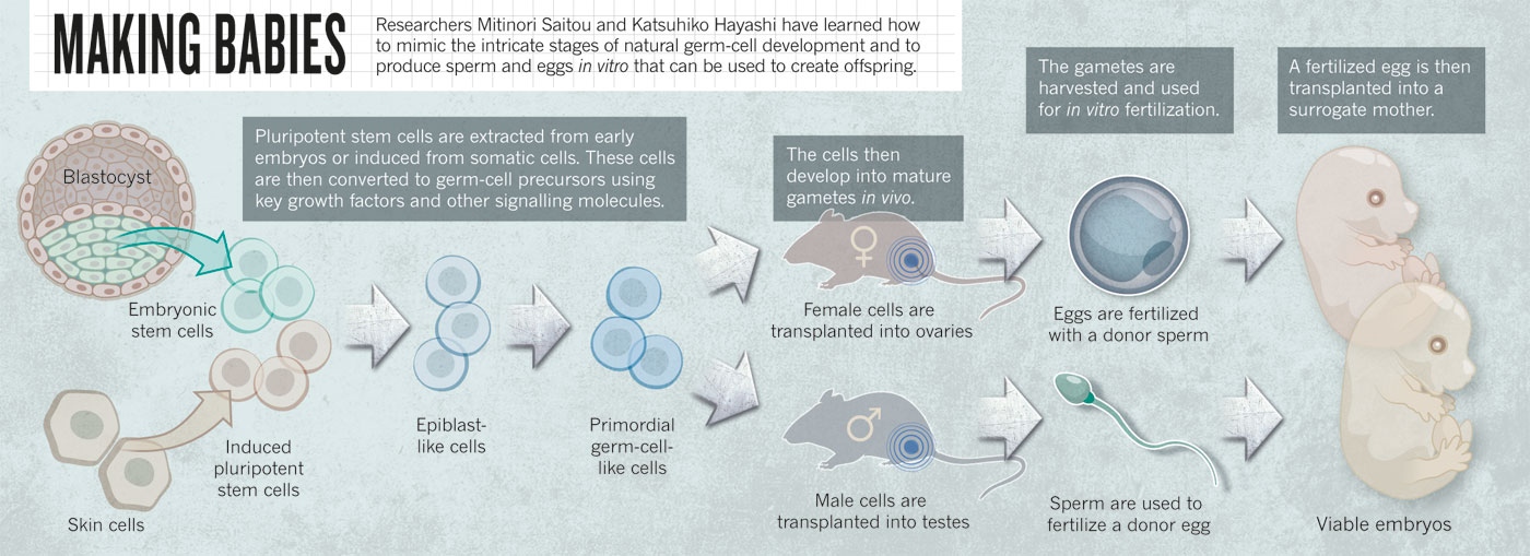
gcs)。为了证明这些实验室培养的原始生殖细胞与自然发育而成的原始生殖细胞类似,他利用它们生成了卵子,进而创造小鼠生命。他表示,这个创造出来的小鼠生命仅仅是他研究的一个“副产品”,他的研究将意味着更多——利用不孕妇女的皮肤细胞为她们提供可受精的卵细胞。与此同时他还提出,男性的皮肤细胞也可以用来创造卵子,同样,女性的皮肤细胞也可以生成精子。(事实上,研究结果发表后,许多同性恋发邮件给Hayashi ,索要更多的信息。)
尽管这是一项创新研究,但是公众的广泛关注还是令Hayashi和他的教授Mitinori Saitou感到非常惊讶。他们花了十多年不断挖掘哺乳动物配子产生的微妙细节,然后在体外条件下重新创建该过程——一切都是为了科研,而非医疗。现在他们的方法使研究人员能够创建无限的原始生殖细胞,这种在以前很难获得的珍贵细胞的正常供应有助于推动哺乳动物生殖研究。
但是,当他们将这个科学挑战自小鼠到猴子,再到人类推进时,这一过程被公众定义为治疗不孕不育的过程,于是相关的道德争议随之出现。“毫无疑问,他们在小鼠身上给这一领域带来了重大的改变,” 洛杉矶加州大学的生育专家Amander Clark说,“但是,在这项技术展示它的实用性之前,我们必须讨论一下使用这种方式创造配子的伦理问题。”
回到最初
在小鼠体内,胚胎发育一周后,便出现约40个左右的原始生殖细胞。这个小小的细胞团进而在雌性小鼠体内形成成千上万的卵细胞,在雄性小鼠体内每天都能生成几百万个精细胞,并能够遗传小鼠的全套遗传信息。Saitou想要了解在这些细胞发育过程中受到了那些信号的控制。
在过去的十年中,Saitou已经通过辛苦研究确定了几个基因——包括Stella, Blimp1 和Prdm14 ——这些基因的某种组合在某些时候对于P
gcs的发育起到了至关重要的作用。利用这些基因作为标记,可以从其他细胞中筛选原始生殖细胞以观察这些细胞的变化。2009年,在日本神户的RIKEN发育生物学中心,他发现,当培养条件适当时,在精确的时间加入骨形态发生蛋白4(BMP4),可以胚胎干细胞转化为原始生殖细胞的。为了验证这一发现,他向胚胎干细胞提供高浓度的BMP4,结果显示,几乎所有的胚胎干细胞都变成了P
gcs。他和科学家们都预计这一过程非常复杂。
![]()
人造小鼠生殖细胞产生小鼠胚胎的过程(点击图片查看大图)
Saitou的方法严格遵循了自然过程,这与其他从事类似研究的人形成了鲜明的对比,以色列魏茨曼科学研究所的干细胞专家Jacob Hanna说。许多科学家尝试通过信号分子轰击干细胞在体外创造特定类型的细胞,然后筛选细胞混合物得到他们想要的细胞。但是他们忽略了这些细胞的自然形成过程和这些人造细胞与自然形成细胞的相似程度。Saitou找出了形成生殖细胞所需的条件,去除多余的信号干扰并将每个过程的时间精确控制,给他的同事们留下了深刻的印象。英国谢菲尔德大学的干细胞生物学家Harry Moore将这种生殖细胞发育的精确重现视为一场“胜利”。
到了2009年, Saitou在小鼠生殖细胞出现之前从外胚层取了一些细胞,这成了研究的起点。但是想要真正掌握这个过程中,Saitou希望从细胞培养开始。
当时正值Hayashi从英国剑桥大学回到日本,和Saitou一样,Hayashi在该领域先驱Azim Surani英国的实验室里完成了4年的研究。Surani盛赞这两位科学家说,他们的“
气质、风格和解决问题的方法能够相互补充”。 Saitou “处理事情时很有系统性、完成目标一心一意”,而Hayashi“工作时更有直觉、视角更广阔、处理问题方法相对更加宽松”,他说。“他们确实形成了一个非常强大的团队。”
Hayashi加入了Saitou京都大学的团队,他很快就发现,那里不同于剑桥。在京都大学,Hayashi用在理论讨论上的时间比曾经少得多,而更多的时间都花在实验上。他说“在日本,我们只管‘做’,这有时是非常低效的,但有时又酝酿着巨大的成功”。
Hayashi同样以外胚层细胞作为起点,但与Saitou不同的是,他试图培养一个能够产生原始生殖细胞的稳定细胞系。可惜这种方法没有奏效。Hayashi借鉴其他研究结果——一个关键调控分子(activin A)和生长因子(bFGF)可以将培养的早期胚胎干细胞转化成类似于外胚层细胞的细胞类型。这引发了Hayashi将这两个因素结合起来的想法,诱导胚胎干细胞分化为外胚层,然后采用Saitou之前的方法把这些细胞成为的P
gcs。通过这种新的方法,他最终获得了成功。
为了证明这些人造的原始生殖细胞是真实的拷贝,他们必须证明这些细胞可以进一步发育成精子和卵子。这一进程是非常复杂和难以理解的。所以研究小组将这一工作留给了自然——Hayashi将P
gcs植入无法产生精子的小鼠的睾丸,观察这些细胞是否会发育。Saitou认为,这是可行的,但还是感到有些担忧。当实验进行到第3或4只小鼠时,他们发现小鼠的输精管里充满了精子。“这一切都发生得恰如其分,我知道他们会产生幼仔,”Hayashi说。研究小组将这些精子注入卵细胞中并植入雌性小鼠的胚胎,结果产生了大量的雌性和雄性后代。
他们利用诱导多能干细胞(iPS)进行反复的实验,成熟的细胞被重新编程为胚胎状态。此外,精子被用于生产幼仔,证明它们具有基本功能——这是干细胞分化领域的罕见成就。Clark说:“这是整个多能性干细胞研究领域里在培养皿中生成全功能细胞类型少有的成功案例之一。”
他们预计形成卵细胞更复杂,但是在去年,Hayashi在体外条件下制作有正常着色的原始生殖细胞并转入白化小鼠的卵巢,将产生的卵细胞体外受精后植入代孕。当透过幼崽半透明的眼睑看到黑色的眼睛时,他知道这一切又成功了。
生殖细胞的回馈
目前,许多研究人员已经能够复制验室培养原始生殖细胞的过程。人造原始生殖细胞特定用于表观遗传学研究:通过修饰DNA确定哪些基因表达。最常见的修饰就是为DNA碱基加上甲基,这些修饰在有些情况下,能够反映生物所经历的历史过程。与其它类型的细胞类似,表观遗传标记改变了原始生殖细胞在胚胎发育过程中的命运,但原始生殖细胞有个与众不同的特点,就是当它们发育成精子和卵子后,表观遗传标记被擦除。这就允许细胞创建能够形成任何类型细胞的受精卵。
表观遗传微妙变化中出现错误将会导致不孕不育并出现器官故障,如如睾丸癌。Surani和Hanna的团队已经利用人造原始生殖细胞研究不同酶在表观遗传调控中的作用,也许有一天,能够解答表观遗传网络如何参与疾病调控。
事实上,体外产生的原始生殖细胞可以为研究提供数百万个细胞,而不是供科学家研究了40个左右,这些细胞可以通过解剖早期胚胎获得。Hanna说:“这是一个大问题,因为我们这里有这些稀有的原始生殖细胞正在经历我们尚不了解的全基因组表观遗传变化。”“体外模型为科学家们提供了前所未有的方便,” Clark表示认同。
临床意义
但是Hayashi和Saitou没有办法向乞求帮助的不孕夫妻提供帮助。在这种方法被运用在临床之前,还有许多问题需要梳理。
Saitou和Hayashi发现,虽然运用他们的技术所产生的后代通常似乎是健康和大量的,但这些后代产生的原始生殖细胞并生不完全“正常”。 第二代原始生殖细胞产生的卵细胞往往是脆弱、畸形的,并且从支持它们生长的组织上脱离。当受精时,卵细胞内部会分为三组染色体,而不是正常的两组,体外受精的成功率也只有正常原始生殖细胞的三分之一。哈佛医学院从事表观遗传学研究的Yi Zhang,使用Saitou的方法在研究中发现,体外受精过程中,人造的原始生殖细胞不能像自然状态下产生的原始生殖细胞一样,抹去它们的表观遗传标记。“我们必须要知道,这些都是P
gcs的类似细胞,而不是真正的原始生殖细胞,”他说。
此外,这项技术还存在两个大的挑战。首先是在不将P
gcs放回睾丸或卵巢的前提下买入和使它们变成成熟的精子和卵子,Hayashi目前正在试图破解P
gcs生成卵子或精子的生物信号,使人工培育条件下完成这一阶段成为可能。
但最可怕的挑战是在人体重复上述所有的工作。该小组已经在利用Saitou找到的关键调控基因来调整人类的iPS细胞,但是Saitou 和Hayashi都知道,人类的信息调控网络不同于小鼠。此外,Saitou有无数的小鼠胚胎进行解剖,但无法在人类胚胎进行相应的实验步骤。幸运的是,他们得到了长达5年共1.2亿日元的资助,每周可以获得20个猴子胚胎。Hayashi表示如果一切顺利的话,他们可以在5-10年内在猴子身上完成所有的工作。随后经过一些小的调整,这种方法就可以用来生产人类原始生殖细胞。
但是利用原始生殖细胞治疗人类不孕不育仍将是一个巨大的跳跃,许多科学家对此持有相当谨慎的态度,这当然包括Saitou在内。iPS和胚胎干细胞在培养过程中常常会出现染色体异常、基因突变等现象。“如果在整个过程中出现一点差错,都可能产生深远的、多代的不良后果。” Moore说。
证明该技术在猴子实验中时安全的,将打消人们的顾虑。但又有新的问题随之而来,要有多少健康的猴子出生,才能证明这个方法是安全的?实验需要观察多少个猴子世代?
最终,还是需要人类胚胎进行测试,而这一进程势必会受到胚胎研究伦理问题的限制。近年来,新的非侵入性成像技术,将帮助医生对胚胎质量的好坏进行精确排序。这种人造胚胎可以利用类似于正常的体外受精胚胎的植入方式进入母体。这些有可能在胚胎研究态度较为宽泛的地区实现。
当技术准备完成,挑战生殖的更大壮举也是有可能实现的。举例来说,人的皮肤细胞理论上可以被用来创造与伴侣精子配对的卵细胞,然后植入子宫孕育。但是存在这样的怀疑,有些事情似乎永远无法实现——国际科学家协会的Hinxton团队认为从男性XY细胞很难获得卵细胞,同理,女性的XX细胞,也很难培养成为精子。
Saitou用雄性小鼠iPS细胞制造出了精子,利用雌性小鼠制造出了卵细胞,他曾表示反过来也是可能的。如果是这样的话,同一只小鼠产生的卵细胞和精子受精,就会产生,以前从未见过的东西:自交小鼠。Hayashi和Saitou都不愿意尝试这个。“如果有充分的科研理由,我们只会在小鼠身上这么做,”Saitou说。但眼下,他还没有看到这样的理由。
这两位科学家已经感受到病人和资金的压力,这项技术可能是无法做试管婴儿的妇女或者是无法产生精子或卵子的人的最后希望。Hayashi表示真正可行的不孕不育治疗方案可能要在10年甚至50年的未来方能实现。“在我看来,那是很遥远的事,我不想给人们不现实的希望,”他说。
许多不孕不育患者只看到了最终的结果——小鼠实验的成功——但却忽略了多年艰苦细致的工作。Hayashi说,他们没有意识到,人类胚胎与小鼠差异非常之大,从小鼠到人类就意味着这一切几乎是从零开始。
参考文献补充文献[img=Non-invasive imaging of human embryos before embryonic genome activation predicts development to the blastocyst stage]http://www.ibioo.com/data/attachment/portal/201308/25/095620zzeozo866db1zeou.gif[/img]
Non-invasive imaging of human embryos before embryonic genome activation predicts development to the blastocyst stage文献检索:
doi:10.1038/nbt.1686We report studies of preimplantation human embryo development that correlate time-lapse image analysis and gene expression profiling. By examining a large set of zygotes from in vitro fertilization (IVF), we find that success in progression to the blastocyst stage can be predicted with >93% sensitivity and specificity by measuring three dynamic, noninvasive imaging parameters by day 2 after fertilization, before embryonic genome activation (EGA). These parameters can be reliably monitored by automated image analysis, confirming that successful development follows a set of carefully orchestrated and predictable events. Moreover, we show that imaging phenotypes reflect molecular programs of the embryo and of individual blastomeres. Single-cell gene expression analysis reveals that blastomeres develop cell autonomously, with some cells advancing to EGA and others arresting. These studies indicate that success and failure in human embryo development is largely determined before EGA. Our methods and algorithms may provide an approach for early diagnosis of embryo potential in assisted reproduction.
[img=Critical function of Prdm14 for the establishment of the germ cell lineage in mice]http://www.ibioo.com/data/attachment/portal/201308/25/095620pwoiyo1b1149bbm1.gif[/img]
Critical function of Prdm14 for the establishment of the germ cell lineage in mice文献检索:
doi:10.1038/ng.186Specification of germ cell fate is fundamental in development and heredity. Recent evidence indicates that in mice, specification of primordial germ cells (P
gcs), the common source of both oocytes and spermatozoa, occurs through the integration of three key events: repression of the somatic program, reacquisition of potential pluripotency and ensuing genome-wide epigenetic reprogramming. Here we provide genetic evidence that Prdm14, a PR domain–containing transcriptional regulator with exclusive expression in the germ cell lineage and pluripotent cell lines, is critical in two of these events, the reacquisition of potential pluripotency and successful epigenetic reprogramming. In Prdm14 mutants, the failure of these two events manifests even in the presence of Prdm1 (also known as Blimp1), a key transcriptional regulator for P
gc specificatio. Our combined evidence demonstrates that Prdm14 defines a previously unknown genetic pathway, initiating independently from Prdm1, for ensuring the launching of the mammalian germ cell lineage.
[img=Blimp1 is a critical determinant of the germ cell lineage in mice]http://www.ibioo.com/data/attachment/portal/201308/25/095620etx0z7oqnx5szhcq.jpg[/img]
Blimp1 is a critical determinant of the germ cell lineage in mice文献检索:
doi:10.1038/nature03813Germ cell fate in mice is induced in pluripotent epiblast cells in response to signals from extraembryonic tissues. The specification of approximately 40 founder primordial germ cells and their segregation from somatic neighbours are important events in early development. We have proposed that a critical event during this specification includes repression of a somatic programme that is adopted by neighbouring cells. Here we show that Blimp1 (also known as Prdm1), a known transcriptional repressor, has a critical role in the foundation of the mouse germ cell lineage, as its disruption causes a block early in the process of primordial germ cell formation. Blimp1-deficient mutant embryos form a tight cluster of about 20 primordial germ cell-like cells, which fail to show the characteristic migration, proliferation and consistent repression of homeobox genes that normally accompany specification of primordial germ cells. Furthermore, our genetic lineage-tracing experiments indicate that the Blimp1-positive cells originating from the proximal posterior epiblast cells are indeed the lineage-restricted primordial germ cell precursors.
[img=A molecular programme for the specification of germ cell fate in mice]http://www.ibioo.com/data/attachment/portal/201308/25/095620y6cam1k6zab1a8cc.jpg[/img]
A molecular programme for the specification of germ cell fate in mice文献检索:
doi:10.1038/nature00927Germ cell fate in mice is induced in proximal epiblast cells by the extra-embryonic ectoderm, and is not acquired through the inheritance of any preformed germ plasm. To determine precisely how germ cells are specified, we performed a genetic screen between single nascent germ cells and their somatic neighbours that share common ancestry. Here we show that fragilis, an interferon-inducible transmembrane protein, marks the onset of germ cell competence, and we propose that through homotypic association, it demarcates germ cells from somatic neighbours. Using single-cell gene expression profiles, we also show that only those cells with the highest expression of fragilis subsequently express stella, a gene that we detected exclusively in lineage-restricted germ cells. The stella positive nascent germ cells exhibit repression of homeobox genes, which may explain their escape from a somatic cell fate and the retention of pluripotency.
[img=Offspring from Oocytes Derived from in Vitro Primordial Germ Cell–like Cells in Mice]http://www.ibioo.com/data/attachment/portal/201308/25/095621zzzvp0b2e0epbrj8.gif[/img]
Offspring from Oocytes Derived from in Vitro Primordial Germ Cell–like Cells in Mice文献检索:
DOI: 10.1126/science.1226889Reconstitution of female germ cell development in vitro is a key challenge in reproductive biology and medicine. We show here that female (XX) embryonic stem cells and induced pluripotent stem cells in mice are induced into primordial germ cell–like cells (P
gcLCs), which, when aggregated with female gonadal somatic cells as reconstituted ovaries, undergo X-reactivation, imprint erasure, and cyst formation, and exhibit meiotic potential. Upon transplantation under mouse ovarian bursa, P
gcLCs in the reconstituted ovaries mature into germinal vesicle-stage oocytes, which then contribute to fertile offspring after in vitro maturation and fertilization. Our culture system serves as a robust foundation for the investigation of key properties of female germ cells, including the acquisition of totipotency, and for the reconstitution of whole female germ cell development in vitro.