主题:【分享】Nano Photos Rival Modern Art--科学与艺术的完美结合

浏览0 回复27 电梯直达
大笨狗
结帖率:
100%
关注:0 |粉丝:0
新手级: 新兵
Every six months, the Materials Research Society celebrates the most eye-catching images found in the course of their researchers' studies -- celebrating the serendipitous convergence of science and art.Materials researchers may struggle for years with stubborn instruments, fragile crystals or difficult chemical reactions before obtaining a bit of precious data from the exotic substances they study. Now, the scrutiny of samples not only yields potentially important data, but also artistic inspiration.

Take a look at the latest finalists.


每隔6个月,材料研究学会即会庆祝他们在研究进程中发现的最引人注目的图片——赞美科学与艺术的完美结合。如今,他们对实验样本的细致分析不仅产生具有潜在重要性的数据,还给人带来颇富美学的灵感。以下是堪比现代艺术品的11幅纳米图片,让人不禁感叹隐藏于科学世界背后的艺术之美。
为您推荐
您可能想找: 气相色谱仪(GC) 询底价
专属顾问快速对接
立即提交
可能感兴趣
大笨狗
结帖率:
100%
关注:0 |粉丝:0
新手级: 新兵
1.金色向日葵

  二氧化硅纳米丝具有令人大为诧异的习惯——能自行组成精美的图画。香港中文大学物理学教授郝少康(S.K. Hark)在扫描电子显微镜下观看二氧化硅纳米丝时,一幅金色向日葵的画面呈现在他的眼前。与植物不同,它们的“肥料”是镓和金催化剂,能让它们长到长度只有几微米,同时直径保持在10纳米左右。郝少康教授给这幅获奖作品着色,令它们与真实的向日葵看上去更加相似。

Silicon oxide nanowires have an amusing habit of arranging themselves into impressive patterns. When S.K. Hark, a professor at the Chinese University of Hong Kong, looked at some of them under a scanning electron microscope, he saw flowers. Unlike plants, their fertilizers were gallium and gold catalysts -- which allowed them to grow to several microns in length while maintaining a roughly 10-nanometer diameter. The physics professor colorized his award-winning crop to enhance their resemblance to real sunflowers.

大笨狗
结帖率:
100%
关注:0 |粉丝:0
新手级: 新兵
2.山川美景

  在新加坡南洋理工大学,博士后学者杨慧英(音译)在检查氧化锌纳米针时,无意中发现了一幅颇为神奇的图片:中国古典油画描绘的山川。为了使这幅图画同真实的山川更为相像,杨慧英给这个场景着色,同时又凭借其独特的想象,增加了一部分的绘画。

At Nanyang Technological University in Singapore, postdoctoral scholar Hui Ying Yang was examining zinc oxide nanoneedles when she saw an image that resembled the mountains as portrayed in classical Chinese paintings. To enhance the resemblance, Yang colorized the scene and added part of the original painting.

大笨狗
结帖率:
100%
关注:0 |粉丝:0
新手级: 新兵
3.山川湖泊与篱笆墙

  美国斯坦福大学鲍哲楠及其研究小组通力合作,使有机晶体管成为更为先进的电子装置。鲍哲楠所指导的研究生刘紫红(音译)用正交极化光显微镜检查这组细微转换。在刘紫红眼中,图片的明亮之处看上去像是山川湖泊,而金电极则像是一堵篱笆墙。

At Stanford University, Zhenan Bao and her team of researchers work to make organic transistors for cutting-edge electronic devices. One of her graduate students, Zihong Liu, used a cross-polarized light microscope to examine this array of the tiny switches. For Liu, bright parts of the film look like lakes and mountains, while the gold electrodes appear to be a fence.


大笨狗
结帖率:
100%
关注:0 |粉丝:0
新手级: 新兵
4.金色森林

  镍钛合金在遭受轮番“虐待”后,跳回“预置”形态,只有经过加热处理,它们才能恢复到原来模式。在德国普朗克冶金研究所,布里特·戈尔·克拉克用聚焦的离子光束以形成这根微柱,接着用纳米刻压机进行压缩。她的传输电子显微镜图片显示了张力对小金属棒的影响。

Nickel-titanium alloys fascinate scientists with their ability to spring back into a pre-set shape after lots of abuse -- just heat them and they will recover. At the Max Planck Institute for Metallurgy in Stuttgart, Germany, Blythe Gore Clark used a focused ion beam to form this micropillar and then a nanoindenter to compress it. Her transmissio-electron-microscope image shows the effects of strain on the tiny metal rod.
大笨狗
结帖率:
100%
关注:0 |粉丝:0
新手级: 新兵
5.大脑横切面
  
    这张聚合体的照片看似肿瘤或大脑的横切面,出自美国伊利诺伊州阿尔贡国家实验室纳米级材料研究中心研究人员穆鲁贾纳汉·拉玛纳汉(Muruganathan Ramanathan)之手。这位博士后学者拍摄了美丽物质聚合体的超薄图片,用活性氧铁蚀刻工艺以图案对其装饰,同时用热量和溶剂使其更加清澈透明。结果,它看上去更像是一幅现代艺术品,似乎与先进的科学研究不沾边。

Looking like a tumor or the cross-section of a brain, this image of a polymer was created by Muruganathan Ramanathan at the Center for Nanoscale Materials at Argonne National Laboratory in Illinois. A postdoctoral scholar, Ramanathan made an ultrathin film of the beautiful substance, patterned it with oxygen-reactive-ion etching and used heat and solvents to make it more crystalline. The result looks more like modern art than the results of a wry scientific study.

大笨狗
结帖率:
100%
关注:0 |粉丝:0
新手级: 新兵
6.向悬崖边奔跑之人

  一天,桑迪亚国家实验室科学家杰弗·布伦尼克将氧化钽晶体插入扫描电子显微镜中,开始搜集图片,他发现这台仪器并没有得到适当地清洁。幸运的是,一些小的聚苯乙烯珠子从上一次实验中遗留下来,粘在实验样本的一侧,形成了一幅令人难以置信的图案,看上去仿佛是一个人向悬崖边奔跑。布伦尼克意识到这幅图案的美术潜力,于是给其着色,提交到材料研究学会,参加“2008年科学与艺术大赛”,最终获得第二名的佳绩。

When Geoff Brennecka, a scientist at Sandia National Laboratory, inserted a tantalum oxide crystal into his scanning electron microscope and started gathering images, he realized the machine had not been cleaned properly. Lucky for him, some tiny polystyrene beads left over from the preceding experiment stuck to the side of his sample in an incredible pattern. It looked like a man running toward the edge of a cliff.

Brennecka saw its artistic potential, colorized the image and then submitted it to the Materials Research Society Spring 2008 Science as Art Competition, where it won second place.


大笨狗
结帖率:
100%
关注:0 |粉丝:0
新手级: 新兵
7.精致悬臂

  这幅几乎完美无暇的金晶体的图片是西班牙马德里大学的维奥利塔·纳瓦罗通过原子力显微镜捕捉的。这些显微镜产生了堪称世界上微小物体的最清晰的图片,极其微小的悬臂在它们的表面回来转动。激光干涉计在悬臂经过原子大小的拐点时,捕捉到它们的轻微活动。

This image of a nearly flawless gold crystal was captured by Violeta Navarro at the University of Madrid with an atomic-force microscope. These microscopes produce some of the most vivid images of tiny objects in the world by running an extremely small cantilever back and forth over their surfaces. Then, laser interferometers pick up slight movements of the cantilever as it passes over atom-sized bumps.


大笨狗
结帖率:
100%
关注:0 |粉丝:0
新手级: 新兵
8.遥远星系

  研究生迈克尔·塞格纳托维茨先是将一些钾铌氧化物堆积到硅表面,接着用光学显微镜拍摄了这幅图片,看上去像一个遥远的星系一般。

After depositing some potassium niobium oxide onto a silicon surface, graduate student Michael Sygnatowicz used an optical microscope to take this photograph, resembling a distant galaxy.


大笨狗
结帖率:
100%
关注:0 |粉丝:0
新手级: 新兵
9.有污迹的窗户

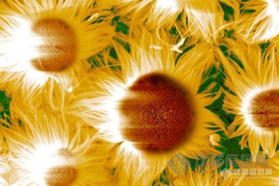
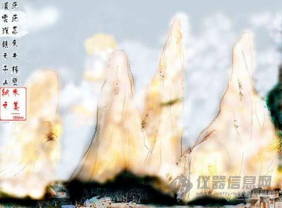
  这幅图片看上去像是一块玻璃有污迹的窗户,但其实它是晶体顶部一层薄薄铁薄膜的磁畴(magnetic domain)。这里的晶体是由镁和镓砷酸盐制成的。意大利ELETTRA同步辐射光源实验室研究人员索利曼·墨索奥尼采用X光线磁循环二色性技术,结合光电子-发射显微镜方法,缔造了这个惊人画面。如果你看到这幅图画时并没有感到丝毫吃惊,那一个更为简单的解释是,墨索奥尼用强大X光线的两个相对偏振分束对其样本进行拍摄,接着从一个个文件上删减数据点。

Although it looks like a stained-glass window, this image shows the magnetic domains of a thin iron film sitting atop a crystal made from magnesium and gallium arsenate. Souliman el Moussaoui, a researcher at the ELETTRA Synchrotron Light Laboratory in Italy, used X-ray magnetic circular dichroism with photoelectron-emission microscopy to create the striking picture. If you didn't pass out trying to read that, a simpler explanation is that el Moussaoui shot the sample with two oppositely polarized beams of powerful X-rays -- and then subtracted the data points in one file from the other.

大笨狗
结帖率:
100%
关注:0 |粉丝:0
新手级: 新兵
10.哈得逊湾森林

  这可能是覆盖在多孔硅模具上的聚合体的扫描电子显微镜图片,对于美国得克萨斯大学的法迪赫·布约克塞林而言,它看上去就像覆盖哈得逊湾的一片森林。

It may be a scanning-electron-microscope image of polymers covering a porous silicon mold, but to researcher Fatih Buyukserin at the University of Texas, it looked like a forest bordering the Hudson River.

猜你喜欢最新推荐热门推荐更多推荐
品牌合作伙伴