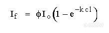
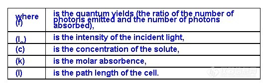
The Single Wavelength Excitation Fluorescence DetectorThe single wavelength excitation fluorescence detector is probably the most sensitive
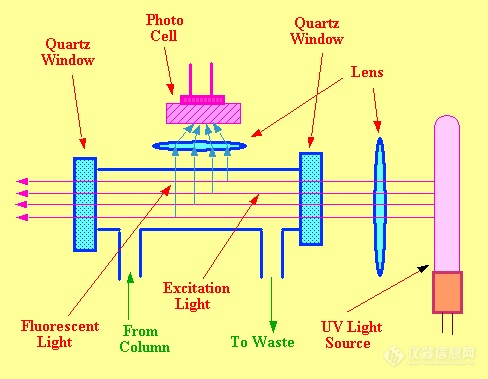
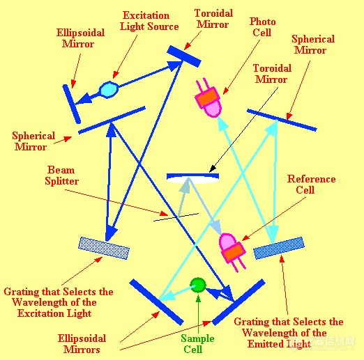
$$lc detector that is available, but is achieved by forfeiting versatility. A diagram of a simple form of the fluorescence detector is shown in figure 36.
The excitation light is normally provided by a low pressure mercury lamp which is comparatively inexpensive and provides relatively high intensity UV light at 253.7 nm. Many substances that fluoresce will be excited by light of this wavelength.
![]() Figure 36. The Single Wavelength Excitation Fluorescent Detector
Figure 36. The Single Wavelength Excitation Fluorescent Detector The excitation light is focused by a quartz lens through the cell. A second lens, set normal to the incident light, focuses the fluorescent light onto a photo cell. A fixed wavelength fluorescence detector will have a sensitivity (minimum detectable concentration at an excitation wavelength of 254 nm) of about 1 x 10⁻⁹ g/ml and a linear dynamic range of about 500 with a response index of 0.96 < r <1.04.